Lakshmi BVS*, Sudhakar M, Anubindu E
Department of Pharmacology, Malla Reddy College of Pharmacy, Dhulapally (via Hakimpet), Maisammaguda, Secunderabad-500014, Telangana, India.
ORIGINAL RESEARCH ARTICLE
Volume 2, Issue 2, Page 2-5, May-August 2014.
Received: 26 July 2014
Revised: 10 August 2014
Accepted: 18 August 2014
Early view: 20 August 2014
mens replica watches Most popular www.beautystic.com article source best replica watches For Sale Online replica shoes.
*Author for correspondence
E-mail: [email protected]
Background: Anti-tumor and antioxidant activity of ethyl acetate extract of Mussaenda philippica sepals (400 mg/kg) was evaluated against Caco-2 cell line in male mice for colon cancer and MCF-7 cell line in female mice for breast cancer.
Materials and methods: After 48 hours of tumor inoculation, the extract was administered daily for 30 days. At the end of the 30 day period, the mice were sacrificed for observation of anti-tumor activity. The effect of Mussaenda philippica sepal extract on the tumor burden, body weight of Caco-2 and MCF-7 bearing hosts and simultaneous alterations in the hematological profile, serum (ALT, LDH, ALP, GGT and glucose) and liver biochemical parameters (lipid peroxidation, GSH, GPx and catalase) were estimated.
Results: The Mussaenda philippica sepal extract showed decrease in abdominal circumference, body weight and organs weight of tumor bearing mice. Hematological profile reverted towards normal levels in extract treated mice. Treatment with Mussaenda philippica sepal extract increased the ferritin levels while CEA (carcinogenic embryonic antigen) levels were found to be decreased. Treatment with Mussaenda philippica sepal extract restored the serum biochemical parameters towards normal levels and decreased the levels of lipid peroxidation and increased the levels of reduced glutathione and other antioxidant enzymes (CAT and GPx).
Conclusion: The ethyl acetate extract of Mussaenda philippica sepal extract exhibited antitumor and antioxidant effect by modulating lipid peroxidation and augmenting antioxidant defense system in Caco-2 and MCF-7 bearing mice.
Keywords: Mussaenda philippica extract, Caco-2, MCF-7, Cell line, Apoptosis, Antitumor,
Antioxidant.
INTRODUCTION
An increasing interest have been attracted for phenolics in recent years because of their significant bioactivities, such as scavenging free radicals, chelating metals, regulating enzyme activity, and modulating cell proliferation (Rice-Evans et al., 1996), which have been associated with the beneficial effects of polyphenol-rich diets on human health. Recent investigations have been shown that antioxidant activities of plant tissues are correlated with oxidative stress defense and different human diseases, including cancer, arteriosclerosis and aging processes (Manosroi et al., 1995). Polyphenolic antioxidants can interfere with the oxidation process by reacting with free radicals, chelating free catalytic metals and acting as oxygen scavengers (Shahidi & Wanasundara, 1992). Thus, antioxidant defensive systems have co-evolved with aerobic metabolism to counteract oxidative damage of reactive oxygen species (ROS). Breast cancer is the most common diagnosed invasive cancer in women and is considered to be one of the leading causes of death due to cancer. Breast cancer is extremely difficult to treat due to several distinct classes of tumors which exhibit different treatment responses.
Epidemiological studies have indicated the relationship between flavonoid intake and reduced risk of certain cancers (Lopez-Otin & Diamandis, 1998). In many studies of dietary prevention of cancer, a model of breast cancer has been established in assessing the impact of a wide variety of flavonoids for their efficacy in inhibiting cancer (Wang et al., 2005). A decreased risk of breast cancer incidence has been associated with a high intake of genistein and moderate consumption of red wine (Tomera, 1999). The biological and pharmacological properties of Chinese herbs have been received more attention in recent years, which is attributed to the fact that Chinese herbs contain a significant amount of phytochemicals, such as antioxidants and other bioactive compounds.
Mussaenda philippica (M. philippica) (Aurorae) of family Rubiaceae is distributed in throughout India, South East Asia. The flowers are small, tube like, expanded into five, ovate lobes, yellowish orange in color. Traditionally the plant is used as dysentery, jaundice, emollient and snake bites. It is reported to have analgesic (Siddique et al., 2007), anticonvulsant (Mates et al., 2000) and antibacterial activities (Abdullah et al., 2012). There are no reports on whether Mussaenda philippica sepal extract has an effect on human cancer-breast cancer and colon cancer cell lines. Considering the rich antioxidant contents of Mussaenda philippica this study investigated possible antitumor effects and antioxidant status of ethyl acetate extract of Mussaenda philippica sepals on the Caco-2 cell line-induced colon cancer and MCF-7 cell line-induced breast cancer in mice.
MATERIALS AND METHODS
Preparation of plant extract: The flowers of Mussaenda philippica were collected from the local regions in Ranga Reddy District, India and authenticated from Department of Botany, Osmania University, Hyderabad, India with voucher no. 01427. Then they were shade dried and grounded coarsely and stored in air tight containers. Coarsely powdered flowers 40 g were packed in a Soxhlet apparatus and extracted using 400 ml ethyl acetate as solvent. After extraction, the extract was obtained by distillation and dried naturally. The extract was then stored at 4 °C in a refrigerator.
Preliminary phytochemical screening
The ethyl acetate extract of Mussaenda philippica was screened for the presence of various phytoconstituents like steroids, alkaloids, glycosides, flavonoids, carbohydrates, amino acids, saponins, terpenoids, tannins and phenolic compounds using the standard procedures.
Determination of LD50 of extract of Mussaenda philippica
Acute toxicity study of Mussaenda philippica sepal extract was carried out for determination of LD50 by adopting fixed dose method of CPCSEA, OECD guideline no. 423. A group of albino mice was used for this study. Acute toxicity studies were conducted and no mortality was observed till the dose of 2000 mg/kg. Hence, 1/5 of the dose 2000 mg/kg i.e. 400 mg/kg has been fixed for the study.
Cell lines
Caco-2 colon cancer cell line and MCF-7 breast cancer cell line: The cell lines were obtained from National Institute of Nutrition, Hyderabad, India. These cells were maintained in bovine serum albumin medium at 37 °C in a humidified atmosphere of 5% CO2 in air.
Animals
Male Wistar albino rats weighing 150-250 g were obtained from National Institute of Nutrition, Hyderabad, India. The rats were housed in polypropylene cages and maintained under standard conditions (12 h light and dark cycles, at 25 ± 3 °C and 35-60% humidity). Standard pelletized feed and tap water were provided ad libitum. All the pharmacological experimental protocols were approved by the Institutional Animal Ethics Committee (Reg no: MRCP/CPCSEA/IAEC/2012-13/MPCOL/03).
Anti-tumor activity of Mussaenda philippica sepal extract against Caco-2 cell line-induced colorectal cancer in Swiss albino mice (Semple et al., 1978)
Twenty four Wistar albino mice were divided into 4 groups (n=6) to study the antioxidant and anti-tumor activity. The animals were grouped as follows:
Group1: Control group (1% tween 80, 1ml)
Group2: Caco-2 cell line induced (0.2ml of 2X10⁶/mouse)
Group3: Caco-2 cell line induced + 5 Fluro uracil (20 mg/kg)
Group4: Caco-2 cell line induced + Mussaenda philippica sepal extract (400 mg/kg body weight).
Anti-tumor activity of Mussaenda philippica sepal extract against MCF-7 cell line-induced breast cancer in Swiss albino mice (Ozello et al., 1960)
Twenty four Wistar albino mice were divided into 4 groups (n=6) to study the antioxidant and anti-tumor activity. The animals were grouped as follows:
Group1: Normal control (saline orally)
Group2: MCF-7 Cell line induced (0.2 ml of 2×10⁶/mouse)
Group3: MCF-7 + 5 Flurouracil (20 mg/kg)
Group 4: MCF-7 Cell line induced + Mussaenda philippica sepal extract (400 mg/kg body weight)
Treatment schedule
Animals were grouped into four groups as explained above. The control group animals were given 1% tween 80- 1ml for 30 days. Group 2 animals were given the respective cancer cell line 2×10⁶ cells/mouse i.p. on the 1st day. Group 3 animals were given respective cancer cell line 2×10⁶ cells/mouse i.p. and 5-flurouracil 20mg/kg body weight i.p. until 30th day respectively. Group 4 animals were given respective cancer cell line 2×10⁶ cells/mouse i.p. on the 1st day and Mussaenda philippica extract 400 mg/kg p.o. from the 3rd day until the 30th day.
Blood sample preparation
The animals were sacrificed on 30th day using ether anesthesia. Blood was collected by carotid bleeding and transferred to anticoagulant EDTA tubes for the estimation of hematological parameters like Hb, RBC and WBC. Some amount of blood was separately collected and was centrifuged using Remi Cool Centrifuge at 4000 rpm for 15 minutes. Serum was separated for the estimation of various biochemical parameters like serum SGPT, alkaline phosphatase, ferritin, carcino embryonic antigen, LDH, GGT and glucose.
Tissue sample preparation
Liver tissue was separated and washed with phosphate buffer saline (0.05 M, pH 7.4). The liver was taken later and minced into small pieces and homogenized in ice cold phosphate buffer saline (0.05 M, pH 7.4) using tissue homogenizer to obtain 1:9 (w/v) (10%) whole homogenate. A part of the liver homogenate was taken and mixed with equal volume of 10% trichloro acetic acid (TCA) for the estimation of malondialdehyde. Homogenate was centrifuged using Remi Cool Centrifuge at 8000 rpm for 30 min. The supernatant was separated and used for estimation of anti-oxidant levels of different enzymes i.e. catalase and reduced glutathione, malondialdehyde and glutathione peroxidase.
Histopathological studies
At the end of the experimental period, the mice were sacrificed, breast and colon were removed. The tissue sample from each group was selected and stored in 10% buffered formalin solution and further embedded in paraffin with wax. The blocks were processed for sectioning; the sections were then stained with haematoxylin and eosin as nuclear and cytoplasmic stains, respectively to assess the activity. Pathological changes, if any, were viewed under light microscope and recorded.
Statistical analysis
The experimental results were expressed as the Mean ± SEM with six rats in each group. Statistical significance of difference between groups was determined by Student’s t-test.
RESULTS
Preliminary phytochemical screening of Mussaenda philippica sepal extract
The main chemical constituents that are found in the sepal extract of Mussaenda philippica include flavonoids, steroids, alkaloids and tannins.
Effect of Mussaenda philippica sepals extract on body weight, organs weight and body circumference in colon cancer induced mice.
The body weight and organs weight of mice from cell line induced group was increased when compared with normal control group. Treatment with the standard drug 5-Fluro uracil showed decrease in body weight and organs weight when compared to the cell line induced animals, whereas treatment with Mussaenda philippica sepal extract showed significant decrease in body weight when compared with the cell line treated animals as mentioned in figure 1-2. The body circumference of cell line induced animals was found to be significantly (P < 0.001) increased than that of the normal control animals. The body circumference in standard and Mussaenda philippica sepal extract group animals was also found to be decreased significantly (P < 0.001) when compared with the cell line induced animals as shown in figure 3.
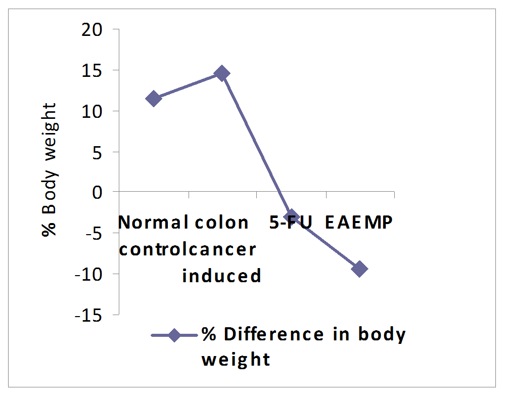 |
Figure 1. Effect of Mussaenda philippica sepal extract on body weight in colon cancer induced mice. 5-FU= 5 Fluro Uracil, EAEMP= Ethyl Acetate Extract of Mussaenda philippica Values are expressed as mean ± SEM. Click here to view full image |
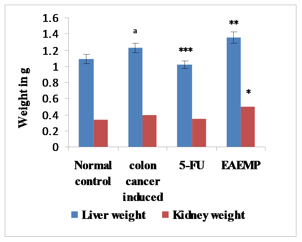 |
Figure 2. Effect of Mussaenda philippica sepal extract on organ weights in colon cancer induced mice. 5-FU= 5 Fluro Uracil, EAEMP= Ethyl Acetate Extract of Mussaenda philippica. Values are expressed as mean ± SEM, (n=6). Click here to view full image |
Data were analyzed by unpaired t test.
***P < 0.001 and *P < 0.05 as compared with cell line induced group, aP < 0.05 as compared with normal control group. Effect of Mussaenda philippica sepal extract on haematological parameters in colon cancer induced mice.
Haematological parameters in cell line induced group were found to be significantly altered compared to those of the normal control group. The leucocytes (WBC) count was found to be increased; RBC and hemoglobin count was significantly decreased in cell line induced animals when compared to the normal control group. Treatment with Mussaenda philippica extract produced a significant increase in Hb level and there was no significant change in RBC count. The WBC levels of Mussaenda philippica treated group were found to be increased when compared to the cell line induced group. Whereas, in the standard treated group with 5-Fluro uracil there was a significant increase in RBC and hemoglobin when compared to the cell line induced animals.
The WBC count was also decreased when compared to the cell line induced animals as shown in table 1.
Effect of Mussaenda philippica sepal extract on serum parameters in colon cancer induced mice.
There was a significant decrease in LDH and glucose levels; significant (P < 0.001) increase in SGPT, GGT and ALP levels in cell line induced animals when compared to the control animals. Whereas, treatment with 5-Flurouracil and Mussaenda philippica sepal extract there was a significant increase in LDH and glucose levels; significant decrease in SGPT, GGT and ALP levels when compared to the cell line induced animals (Table 2).
Effect of Mussaenda philippica sepal extract on catalase, MDA, GSH and GPx in colon cancer induced mice.
Catalase, GSH, and GPx were significantly (P < 0.001) decreased and MDA levels were significantly (P < 0.001) increased in the colon cancer group when compared to the normal group. Treatment with 5 fluorouracil and Mussaenda philippica sepal extract significantly (P < 0.001) decreased the MDA levels and increased the Catalase, GSH, and GPx levels towards the normal (Table 3).
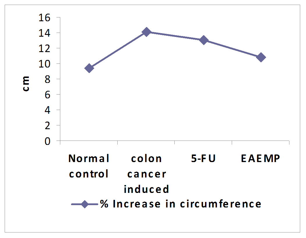 |
Figure 3. Effect of Mussaenda philippica sepal extract on %increase in circumference in colon cancer induced mice. FU= 5 Fluro Uracil, EAEMP= Ethyl Acetate Extract of Mussaenda philippica. Values are expressed as mean ± SEM, (n=6). Click here to view full image |
 |
Table1. Effect of extract of Mussaenda philippica sepal extract on hematological estimations of colon cancer induced mice. Click here to view full image |
5-FU= 5 Fluro Uracil, EAEMP= Ethyl Acetate Extract of Mussaenda philippica.
Values are expressed as mean ± SEM., Data was analyzed by unpaired t test.
cP < 0.001, bP < 0.01 and aP < 0.05 as compared with the normal control; ***P < 0.001 and **P < 0.01 as compared with cell line induced group.
 |
Table 02. Effect of Mussaenda philippica sepal extract on serum biochemical parameters in colon cancer induced mice. Click here to view full image |
5-FU= 5 Fluro Uracil, EAEMP= Ethyl Acetate Extract of Mussaenda philippica.
Values are expressed as mean ± SEM, (n=6). Data were analyzed by unpaired t test.
cP < 0.001 and bP < 0.01as compared with the normal control; ***P < 0.001 and **P < 0.01 as compared with cell line induced group.
 |
Table 3. Effect of Mussaenda philippica sepal extract on catalase, MDA, GSH and GPX in colon cancer induced mice. Click here to view full image |
5-FU= 5 Fluro Uracil, EAEMP= Ethyl Acetate Extract of Mussaenda philippica.
Values are expressed as mean ± SEM, (n=6). Data was analyzed by unpaired t test.
cP < 0.001 as compared with the normal control; ***P < 0.001 **P < 0.01and *P < 0.05 as compared with cell line induced group. Effect of Mussaenda philippica extract on ferritin and carcino embryonic antigen levels in colon cancer induced mice.
Ferritin levels were significantly (P < 0.001) decreased in the colon cancer induced group compared to the normal control group. Treatment with 5 fluorouracil and Mussaenda philippica sepal extract significantly (P < 0.001) restored the levels of ferritin towards the normal. The levels of carcino embryonic antigen were significantly increased in the colon cancer induced group compared to the normal control group. Treatment with 5 fluorouracil and Mussaenda philippica extract significantly (P < 0.001) restored the levels of carcino embryonic antigen towards the normal (Fig. 4).
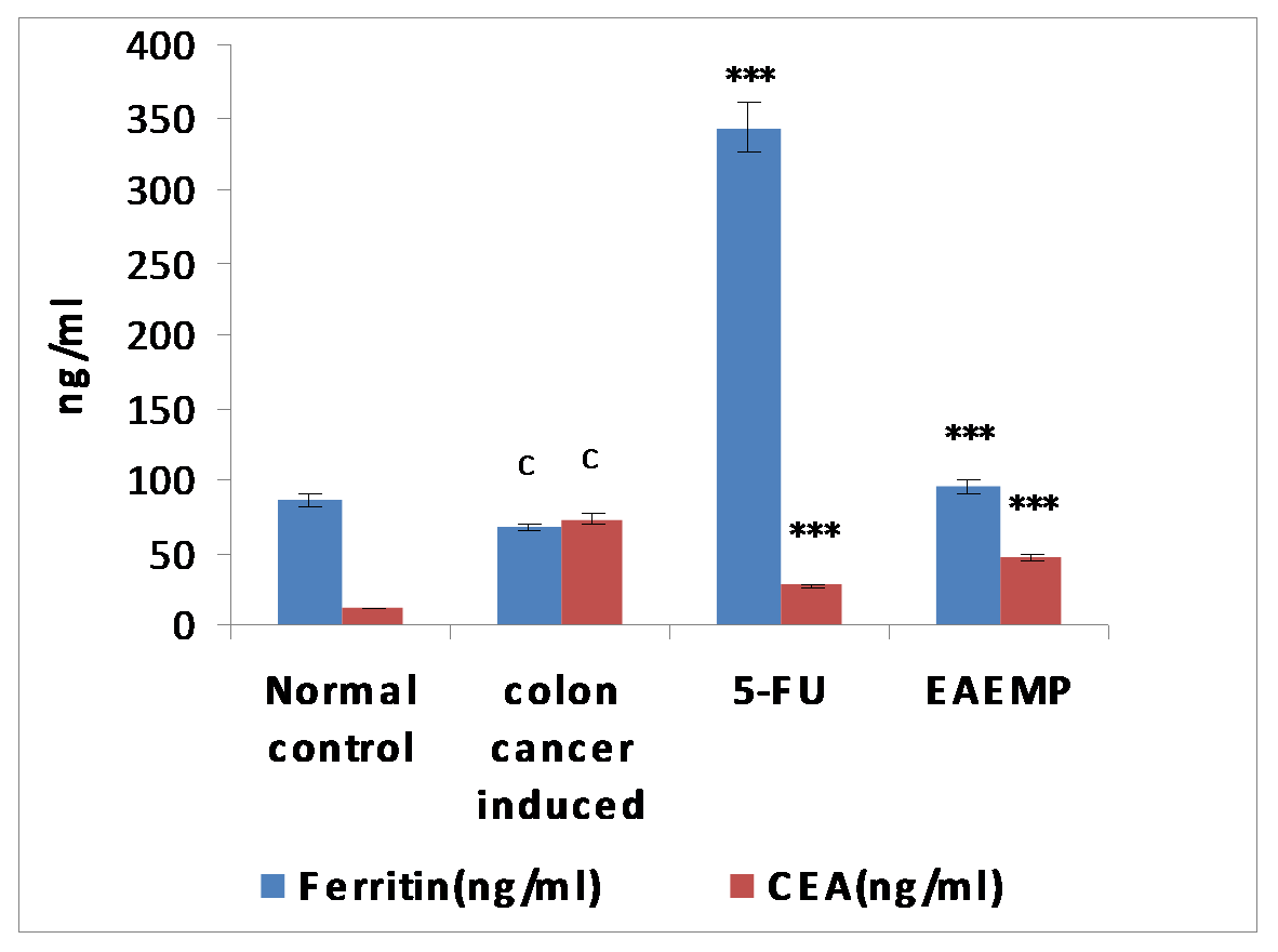 |
Figure 4. Effect of Mussaenda philippica sepal extract on ferritin and carcinoembryonic antigen (CEA) on Caco-2 cell line-induced mice. 5-FU= 5 Fluro Uracil, EAEMP= Ethyl Acetate Extract of Mussaenda philippica. Values are expressed as mean ± SEM, (n=6). Click here to view full image |
Data were analyzed by unpaired t test.
cP < 0.001 as compared with the normal control; ***P < 0.001 as compared with cell line induced group. Effect of Mussaenda philippica sepal extract on body weight, organs weight and body circumference in breast cancer induced mice.
The body weight and organs weight of mice from cell line induced group was increased when compared with normal control group. Treatment with the standard drug 5-Fluro uracil showed decrease in body weight and organs weight when compared to the cell line induced animals, whereas treatment with Mussaenda philippica sepal extract showed significant decrease in body weight when compared with the cell line treated animals as mentioned in figure 5-6. The body circumference of cell line induced animals was found to be significantly (P < 0.001) increased than that of the normal control animals. The body circumference in standard and Mussaenda philippica sepal extract group animals was also found to be decreased significantly (P < 0.001) when compared with the cell line induced animals as shown in figure 7.
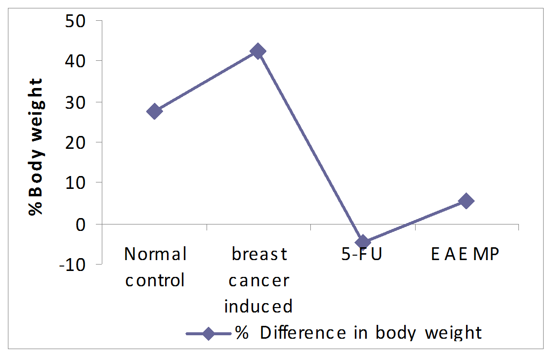 |
Figure 5. Effect of Mussaenda philippica sepal extract on body weight in breast cancer induced mice. 5-FU= 5 Fluro Uracil, EAEMP= Ethyl Acetate Extract of Mussaenda philippica. Click here to view full image |
Effect of Mussaenda philippica sepal extract on haematological parameters in breast cancer induced mice.
Haematological parameters in cell line induced group were found to be significantly altered compared to those of the normal control group. The leucocytes (WBC) count was found to be increased; RBC and hemoglobin count was significantly decreased in cell line induced animals when compared to the normal control group. Treatment with Mussaenda philippica extract produced a significant increase in Hb level and there was no significant change in RBC count. The WBC levels of Mussaenda philippica treated group were found to be increased when compared to the cell line induced group. Whereas, in the standard treated group with 5-fluro uracil there was a significant increase in RBC and hemoglobin when compared to the cell line induced animals. The WBC count was also decreased when compared to the cell line induced animals as shown in table 4.
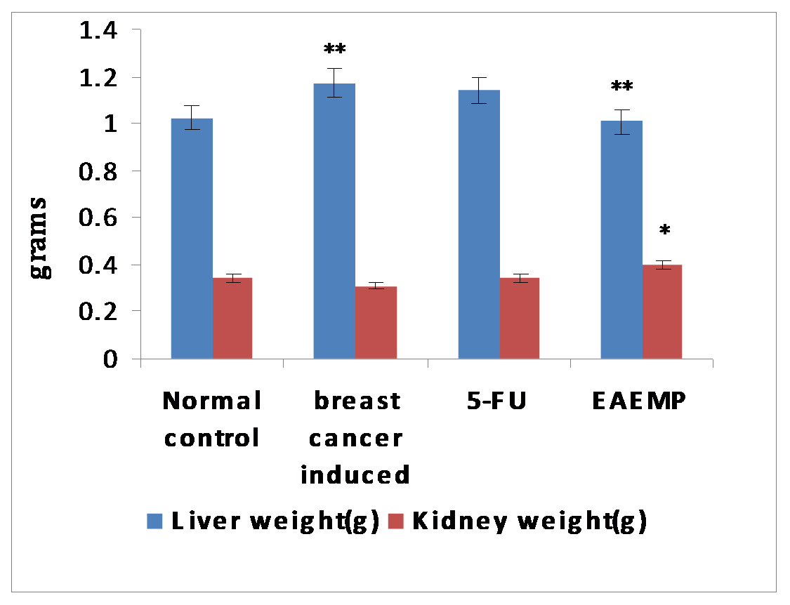 |
Figure 6. Effect of Mussaenda philippica sepal extract on organ weights. 5-FU= 5 Fluro Uracil, EAEMP= Ethyl Acetate Extract of Mussaenda philippica. Values are expressed as mean ± SEM, (n=6). Click here to view full image |
Data was analyzed by unpaired t test.
aP < 0.01 when compared to the normal control. ***P < 0.001 and *P < 0.05 as compared with cell line induced group.
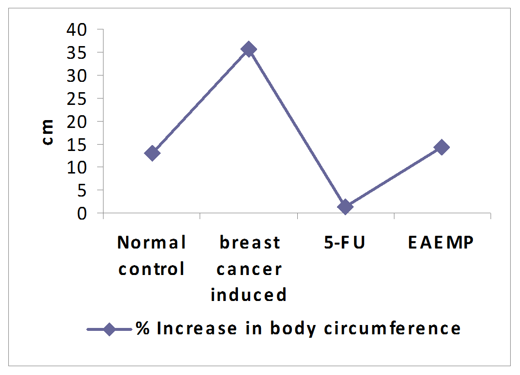 |
Figure 7. Effect of Mussaenda philippica sepal extract on body circumference in breast cancer induced mice. 5-FU= 5 Fluro Uracil, EAEMP= Ethyl Acetate Extract of Mussaenda philippica. Values are expressed as mean ± SEM, (n=6). Click here to view full image |
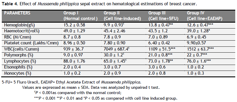 |
Table 4. Effect of Mussaenda philippica sepal extract on hematological estimations of breast cancer. Click here to view full image |
5-FU= 5 Fluro Uracil, EAEMP= Ethyl Acetate Extract of Mussaenda philippica.
Values are expressed as mean ± SEM. Data was analyzed by unpaired t test.
cP < 0.001as compared with the normal control; ***P < 0.001 **P < 0.01 and *P < 0.05 as compared with cell line induced group.
 |
Table 5. Effect of Mussaenda philippica sepal extract on serum biochemical parameters in breast cancer induced mice. Click here to view full image |
5-FU= 5 Fluro Uracil, EAEMP= Ethyl Acetate Extract of Mussaenda philippica.
Values are expressed as mean ± SEM, (n=6). Data were analyzed by unpaired t test.
c P < 0.001, bP < 0.01 and as compared with the normal control; ***P < 0.001 **P < 0.01and *P < 0.05 as compared with cell line induced group. Effect of Mussaenda philippica sepal extract on catalase, MDA, GSH and GPx in breast cancer induced mice.
Catalase, GSH, and GPx were significantly (P < 0.001) decreased and MDA levels were significantly (P < 0.001) increased in the breast cancer group when compared to the normal group. Treatment with 5 fluorouracil and Mussaenda philippica sepal extract significantly (P < 0.001) decreased the MDA levels and increased the Catalase, GSH, and GPx levels towards the normal (Table 6).
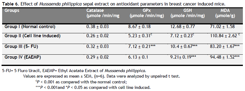 |
Table 6. Effect of Mussaenda philippica sepal extract on antioxidant parameters in breast cancer induced mice. Click here to view full image |
5-FU= 5 Fluro Uracil, EAEMP= Ethyl Acetate Extract of Mussaenda philippica.
Values are expressed as mean ± SEM, (n=6). Data were analyzed by unpaired t test.
cP < 0.001 as compared with the normal control; ***P < 0.001and *P < 0.05 as compared with cell line induced. Effect of Mussaenda philippica extract on ferritin and carcino embryonic antigen levels in breast cancer induced mice.
Ferritin levels were significantly (P < 0.001) decreased in the breast cancer induced group compared to the normal control group. Treatment with 5 fluorouracil and Mussaenda philippica sepal extract significantly (P < 0.001) restored the levels of ferritin towards the normal. The levels of Carcino embryonic antigen were significantly increased in the breast cancer induced group compared to the normal control group. Treatment with 5 fluorouracil and Mussaenda philippica extract significantly (P < 0.001) restored the levels of Carcino embryonic antigen towards the normal (Figure 8).
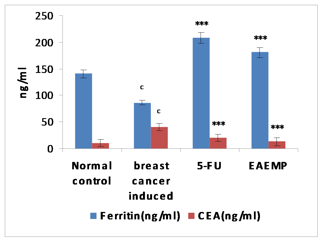 |
Figure 8. Effect of Mussaenda philippica sepal extract on Ferritin and carcinoembryonic antigen (CEA) in breast cancer induced mice. 5-FU= 5 Fluro Uracil, EAEMP= Ethyl Acetate Extract of Mussaenda philippica. Values are expressed as mean ± SEM, (n=6). Click here to view full image |
Data was analyzed by unpaired t test.
cP < 0.001as compared with the normal control; ***P < 0.001 as compared with cell line induced group. DISCUSSION
Plants have the ability to synthesize a wide variety of chemical compounds that are used to perform important biological functions, and to defend against attack from predators such as insects, fungi and herbivorous mammals. Many of these phytochemicals have beneficial effects on long-term health when consumed by humans, and can be used to effectively treat human diseases. Cancer is a worldwide disease with an estimated one million new cases and a half million deaths each year.
Histopathological estimation
Histopathology of colon
 |
Fig. 9 (a) Normal control Yellow arrow indicates the mucosal region of colon which appeared normal. The red arrow indicates the sub mucosal region containing mucosal glands which appeared normal. The white arrow indicates the muscular region which appeared normal as well. Tissue is stained with Haematoxylin and Eosin at magnification 40X. Click here to view full image |
The present study was carried out to evaluate antioxidant & anticancer activity of ethyl acetate extract of Mussaenda philippica sepals on cell line induced in vivo breast cancer and colon cancer. Chemoprevention involves the use of either natural or synthetic compounds to delay inhibit or reverse the development of cancer in normal or pre-neoplastic conditions. In the present study the anticancer activity was evaluated against two specific organ cancers i.e., colon cancer and breast cancer. This cancer was induced by specific cancer cell lines. The Mussaenda philippica sepal extract treated animals (400 mg/kg) significantly brought back the hematological parameters to more or less normal levels.
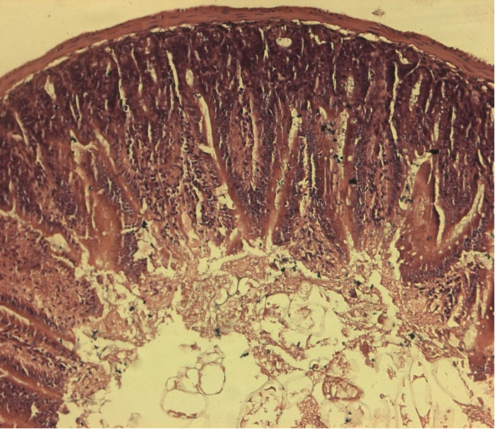 |
Fig. 9 (b) Cell line induced: Red arrow indicates the mucosal degeneration of intestinal epithelial cells in the mucosal layer. The white arrow indicates the sub mucosal glands and the yellow arrow indicates muscular region which appeared normal. Tissue is stained with Haematoxylin and Eosin at magnification 40X. Click here to view full image |
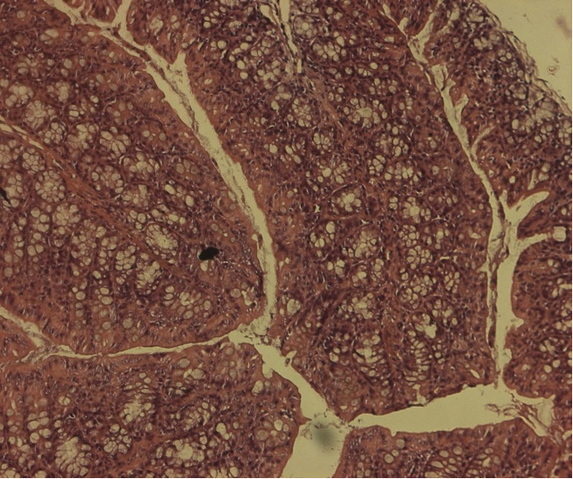 |
Fig. 9 (c) 5-FU: Yellow color indicates goblet cells proliferation in the mucosal and sub mucosal glandular region. Tissue is stained with Haematoxylin and Eosin at magnification 40X. Click here to view full image |
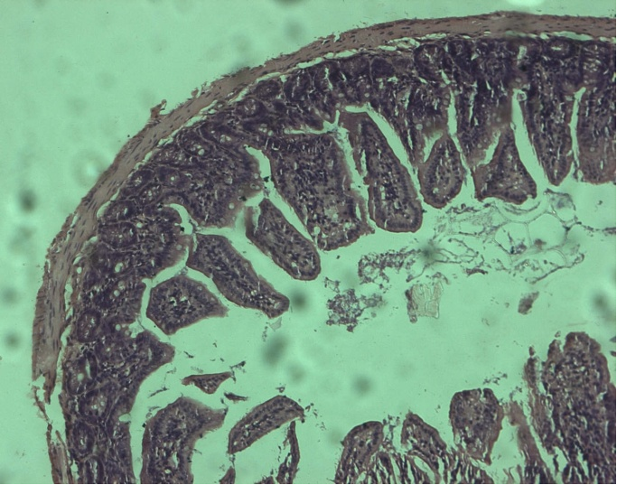 |
Fig. 9 (d) EAEMP: Yellow color indicates the mucosal epithelium which appeared normal. Sub mucosal glands appeared normal which is indicated by the white arrow. Click here to view full image |
Histopathology of breast tissue
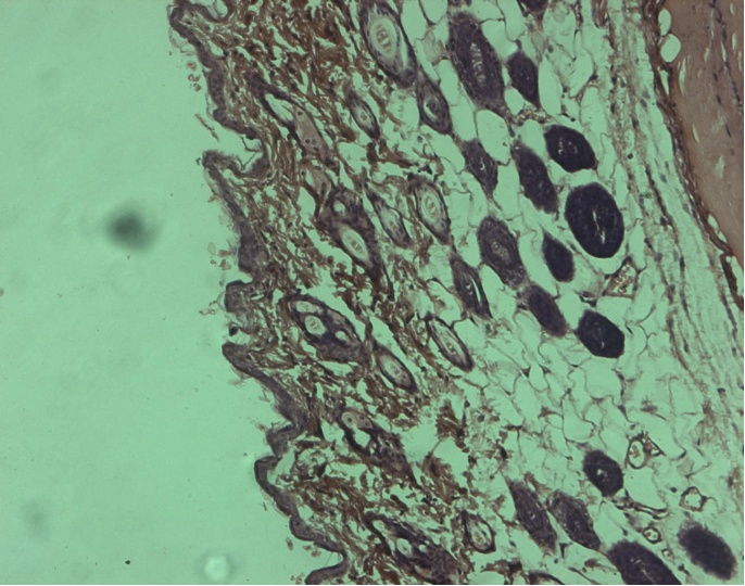 |
Fig. 10 (a) Normal control: Epidermis appeared normal which is indicated by the white arrow. The yellow color indicates dermis and connective tissue which appeared normal. Hair follicles appeared normal which is indicated by the red arrow. Tissue is stained with Haematoxylin and Eosin at magnification 40X. Click here to view full image |
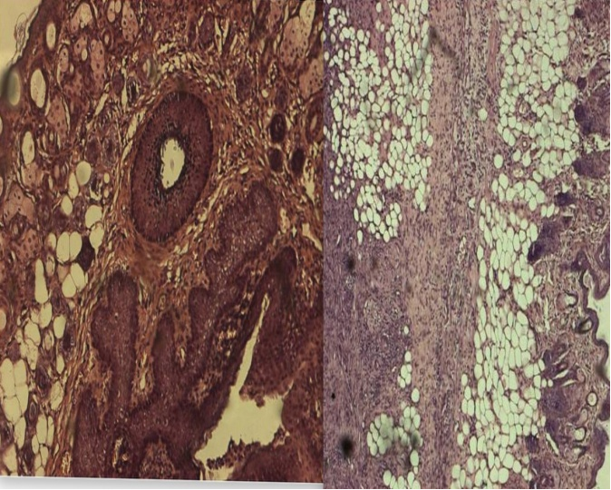 |
Fig. 10 (b) Cell line-induced: Red arrow indicates proliferation of sebaceous gland. White arrow indicates epithelial cell hyperplasia in the hair follicle. Severe sub mucosal inflammation and fibrosis noticed. Infiltration of inflammatory cells [lymphocytes, neutrophils] noticed and which is surrounded by thick layer of connective tissue which is surrounded by thick layer of connective tissue formation leads to fibrosis (green arrow). Tissue is stained with Haematoxylin and Eosin at magnification 40X. Click here to view full image |
In acute toxicity studies, the administration of Mussaenda philippica sepal extract at the dose of 50, 50, 300, and 2000 mg/kg did not exhibit any adverse effect.
Usually, in cancer chemotherapy the major problems that are being encountered are of myelosuppression and anemia. The anemia encountered in tumor bearing mice is mainly due to reduction in RBC or hemoglobin percentage, and this may occur either due to iron deficiency or due to hemolytic or myelopathic conditions. Treatment with Mussaenda philippica sepal extract brought back the hemoglobin content, RBC, and WBC count more or less to normal levels. This indicates that Mussaenda philippica sepal extract possess protective action on the hemopoietic system (Naveena et al., 2011).
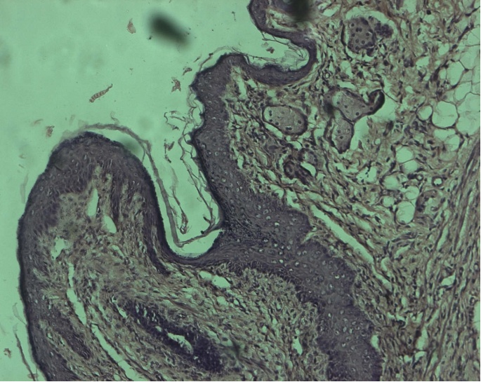 |
Fig. 10 (c) 5-FU: The yellow arrow indicates hyperplasia of epidermal layer. Particularly stratum basal epithelial layer showed continuous proliferation and hyperplastic changes. Dermis, hair follicle and sebaceous gland appeared normal. Tissue is stained with Haematoxylin and Eosin at magnification 40X. Click here to view full image |
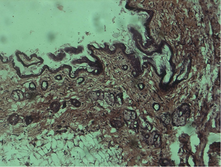 |
Fig. 10 (d) EAEMP: White arrow indicates epidermis which appeared normal. The red arrow indicates dermis, subcutaneous and sebaceous glands and yellow arrow which indicates the hair follicle appeared normal. Tissue is stained with Haematoxylin and Eosin at magnification 40X. In the present experimental work, after the tumor transplantation there was increase in body weight in cancer induced mice due to increase multiplication and uncontrolled growth of cells. The cell line induced mice showed significant increase in body weight. The significant normal body weight was maintained or recovered in animals treated with standard (5-florouracil) and ethyl acetate extract of Mussaenda philippica in the study when compared to cell induced group in both breast cancer and colon cancer. Click here to view full image |
Measurement of serum ferritin is made primarily to assess the level of body iron stores. Serum ferritin levels correlate well with other direct measurements of iron stores like liver biopsy and microscopic examination of bone marrow aspirates for stainable iron deposits. Ferritin is the major intracellular iron storage protein in all organisms (Kishida et al., 1994). Iron serves as a nutrient for cell growth. Iron is an absolute requirement for cell proliferation, as iron-containing proteins catalyze key reactions involved in oxygen sensing, energy metabolism, respiration and DNA synthesis. The ferritin levels were found to be increased in the extract treated group when compared to the cell line induced group.
Carcinoembryonic antigen (CEA) is a glycoprotein involved in cell adhesion. An increased level of CEA was observed in colorectal cancer and breast cancer. CEA measurement is mainly used as a tumor marker to monitor cancer treatment. In the present study increased levels of CEA were observed in cell induced mice in both breast cancer and colon cancer when compared to normal control mice. The Mussaenda philippica animals treated with extract showed decreased levels of CEA in the serum when compared to cell line induced groups. The levels of SGPT and ALP were significantly (P < 0.001) changed in colon and breast cancer induced groups compared to their respective controls and these levels were restored in the Mussaenda philippica sepal extract treated groups.
LDH (lactate dehydrogenase) is released into the serum due to cell membrane damage. LDH catalyzes the oxidation of lactate into pyruvate in the presence of NAD which is subsequently reduced to NADH (Khan et al., 1991). In cell line induced groups of breast cancer and colon cancer the increase in LDH is due to ROS (reactive oxygen species) and the Mussaenda philippica extract treated groups showed significant decrease in LDH levels.
Gamma glutamyl transferase (GGT) is an enzyme found mainly in serum from hepatic origin. GGT catalyzes the degradation of glutathione. Elevated levels are found in hepatobiliary and pancreatic diseases, chronic alcoholism, myocardial infarction with secondary liver damage and diabetes (Dearnley et al., 1981). A dysregulated expression of GGT has been detected in several tumor types like colon, liver, breast. In the present study the cell line induced mice in breast cancer and colon cancer showed significant increase in levels of GGT when compared to normal control group. The Mussaenda philippica extract treated group in both the cancers maintained the levels of GGT.
Glucose is the main source of energy for the cells to grow. The breakdown of glucose to provide energy is called glycolysis. Healthy cells can use other forms of ´food´ like fats, as precursors but cancer cells cannot. Cancer cells need supplies of common glucose to grow. They can only derive energy from glycolysis in the cytoplasm for which they use stored glucose in the body thereby decreasing levels of glucose in the serum. In the present study the decreased levels of serum glucose were observed in cell line induced mice when compared to normal control group. The Mussaenda philippica animals treated with extract restored or maintained the glucose levels in the serum as normal when compared to cell line induced groups.
The extract also restored the hepatic lipid peroxidation and free radical scavenging agent GSH as well as antioxidant enzymes such as MDA and CAT in tumor-bearing mice to near normal levels. Lipid peroxidation, an autocatalytic free radical chain propagating reaction, is known to be associated with pathological conditions of a cell. Malondialdehyde (MDA), the end product of lipid peroxidation, was reported to be higher in cancer tissues than in non diseased organ.
The free radical hypothesis supported the fact that the antioxidants effectively inhibit the tumor, and the observed properties may be attributed to the antioxidant and antitumor principles present in the extract. The preliminary phytochemical screening indicated the presence of alkaloids and flavonoids in Mussaenda philippica. Flavonoids have been shown to possess antimutagenic and antimalignant effects. Moreover, flavonoids have a chemopreventive role in cancer through their effects on signal transduction in cell proliferation and angiogenesis. The cytotoxic and antitumor properties of the extract may be due to these compounds.
The above parameters were supported by the histopathological studies (Fig. 9-10) and these could be responsible for the antitumor and antioxidant activities of Mussaenda philippica.
CONCLUSION
To the best of our knowledge, this is the first study to demonstrate that Mussaenda philippica extract exert significant anti-proliferative and antioxidant against Caco-2 cells in a concentration and time dependent manner. Thus, it can be concluded that Mussaenda philippica extract might have therapeutic value against colon and breast cancer. However, further investigations on a cellular or molecular level are necessary to describe possible mechanism(s) that cause these effects of Mussaenda philippica extract.
ACKNOWLEDGEMENTS
The authors are thankful to the authorities of Malla Reddy College of Pharmacy, Secunderabad, TS, for providing support to the present study.
CONFLICT OF INTEREST
None declared.
REFERENCES
Abdullah E, Raus RA, Jamal P. Evaluation of anti-bacterial activity of Mussaenda philippica to inhibit Bacillus subtilis and Escherichia coli. American Med J 3, 27-32, 2012.
Dearnaley DP, Patel S, Powles TJ, Coombes RC. Carcinoembryonic antigen estimation in cerebrospinal fluid in patients with metastatic breast cancer. Oncodev Biol Med. 2, 305-11, 1981.
Khan N, Tyagi SP, Salahuddin. Diagnostic and prognostic significance of serum cholinesterase and lactate dehydrogenase in Breast cancer. Indian J Pathol Microbiol 34,126-30, 1991.
Kishida T1, Sato J, Fujimori S, Minami S, Yamakado S, Tamagawa Y, Taguchi F, Yoshida Y, Kobayashi M. Clinical significance of serum iron and ferritin in patients with colorectal cancer. J Gastroenterol. 29, 19-23, 1994.
Lopez-Otin C, Diamandis EP. Breast cancer and prostate cancer: an analysis of common epidemiological, genetic, and biochemical features. Endocrine Rev. 19, 365-396, 1998.
Manosroi A, Miller NJ, Bolwell PG, Bramley PM, Pridham JB. The relative antioxidant activity of plant derived polyphenolic flavonoids. Free Rad Res. 22, 375-383, 1995.
Mates MJ, Jimenez S, Fransisca. Evaluation of anti-convulsant activity of leaves and sepals of Mussaenda philippica. Asian Pac J Trop Biomed. C-576, 32, 157,170, 2000.
Naveena, Harath BK, Selvasubramanian. Antitumor activity of Aloevera against Ehrlich ascites carcinoma (EAC) in Swiss albino mice. Int J Pharm Biol Sci. 2, 40-50, 2011.
Ozello L, Lasfargues EY, Murray MR. Growth-promoting activity of acid mucopolysaccharides on a strain of human mammary carcinoma cells. Cancer Res. 20, 600-604, 1960.
Rice-Evans CA, Miller NJ, Paganga G. Structure–antioxidant activity relationship of flavonoids and phenolic acid. Free Rad Biol Med. 20, 7933-7956, 1996.
Semple TU, Quinn LA, Woods LK, Moore GE. Tumor and lymphoid cell lines from a patient with carcinoma of the colon for a cytotoxicity model. Cancer Res. 38, 1345-55, 1978.
Shahidi F, Wanasundara PKJPD. Phenolic antioxidants. Crit Rev Food Sci. 32, 67-103, 1992.
Siddique AB, Sikder MAA, Rashid RB, Islam F, Hossian AKMN. Screening of Analgesic activity for Mussaenda philippica on Swiss-albino mice. Medscape Gen Med. 9, 60, 2007.
Tomera JF. Current knowledge of the health benefits and disadvantages of wine consumption. Trends Food Sci Tech. 10, 129-138, 1999.
Wang Y, Chan FL, Chen S, Leung LK. The plant polyphenol butein inhibits testosterone-induced proliferation in breast cancer cells expressing aromatase. Life Sci. 77, 39-51, 2005.


 This work is licensed under a Creative Commons Attribution-NonCommercial-ShareAlike 4.0 International License.